This article is part one of a two-part series on radiation therapy. Read about external beam radiation here in part two to see how these complementary techniques are used in cancer care.
Hey Doc – one of my buddies at work has cancer and his oncologist recommended radiation therapy. Can you tell me how that works? Thanks!
You know, there’s a simple answer and a long answer to your question; the simple answer is so short I’ll start with that – radiation “burns out” the cancer by killing the cancer cells. But that’s sort of unsatisfying, especially since there are so many types of radiation therapy. So let me go on to the longer answer to fill in some of the details. And there are a lot of them – this might run to two posts…but let me get started and see how it goes.
Radiation therapy goes back to the very earliest days of radiation science – less than three years after Roentgen and Becquerel discovered radiation and radioactivity (respectively) doctors were learning to use radiation to fight cancer. Early work used x-rays to try to destroy cancers directly as well as inserting small metal capsules filled with radium or radon into the tumor to irradiate and kill the cancer cells. These were both reasonably effective – but they were also limited; it was hard to treat blood cancers this way, for example. With time, other nuclides were produced and put to use, engineers found better ways to produce beams of radiation and then learned to shape them to conform to the shape of the tumor (sparing healthy tissue), learned to focus the beams on the tumor to give still lower dose to the surrounding healthy tissues, and even found new forms of radiation to use – we’ll get into all of these.
The process
If a person is diagnosed with cancer they’ll sit down with one of more physicians to decide which form of therapy to use – the main options are surgery, medication (chemotherapy), or radiation; sometimes, owing to its location, the type of cancer, the patient’s overall health, or any of a number of other factors the doctors might feel the cancer is more amenable to one type of treatment or perhaps a combination of treatments (e.g. surgery and chemo); after considering the options the patient and physicians will settle on a treatment. If radiation is part of the treatment plan, then the radiation oncologist and medical physicist are put to work.
The next decision is how the radiation should be applied (more on this below) and then the oncology team will plan the exposure – how much dose will be required to destroy the cancer, how best to get that dose to the tumor, and how to destroy the tumor with the least damage to healthy tissues. This will often involve “fractionation” – delivering the radiation over the course of multiple exposures rather than as a single massive dose to minimize damage to the healthy cells surrounding a tumor while destroying the cancer cells; this is the reason, too, for delivering dose from multiple angles so that, while the tumor is always irradiated, the healthy tissues through which the radiation beam passes will change from fraction to fraction. This, too, is a part of the treatment planning. After the planning comes the irradiation.
Types of radiation therapy
Radiation therapy falls into two primary categories – in one category the cancer is treated by aiming a beam of radiation at it from the outside of the body; that beam can come from an x-ray machine, a particle accelerator, or from a radioactive source and it can consist of high-energy photons (e.g. x-rays or gamma rays) or particles (protons, for example). In the second category, radioactive sources are inserted into the tumor permanently or temporarily, irradiating it from within to destroy the cancerous tissue. Inserting radioactive sources into the tumor is called brachytherapy – let’s start with that.
Brachytherapy
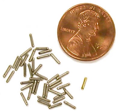
“Brachy” is Greek for “short” and, in brachytherapy, the sources are placed at a short distance from the tumor – in most cases the sources are actually placed inside the tumor itself. Brachytherapy itself is broken into two primary categories – LDR (low dose rate) and HDR (high dose rate) brachytherapy using, as you might guess, sources with less or more radioactivity. I’ve provided radiological support for both in the form of performing radiation surveys and, for HDR, installing temporary radiation shielding to protect visitors and medical staff from the radiation emitted by the sources…I’ll get to that in a little bit.
What I’m most familiar with is the LDR brachytherapy – inserting dozens or even a few hundred radioactive “seeds” into a tumor in a carefully determined three-dimensional pattern designed to provide a high and relatively even radiation field that drops off quickly beyond the edge of the tumor to spare as much healthy tissue as possible. Twenty years ago, when I was covering these procedures, a medical physicist would be plotting the placement of the seeds and observing the resulting dose contours as the seeds were being implanted.
The implantation procedure was pretty straightforward – seeds were loaded into a long needle that was inserted into the tumor and were injected at carefully calculated locations to give the desired radiation dose distribution. Over the next days to weeks or months (depending on the nuclide used) the sources continued irradiating the tumor until they finally decay to stability. Short-lived nuclides can be used to treat an aggressive tumor, producing a high total dose in a relatively short period of time. For less aggressive tumors, a longer-lived nuclide will produce lower radiation levels, reducing the dose rate to family members, coworkers, and others while still successfully treating the cancer.
And my part? I made radiation measurements during the procedure but, mostly, I made sure that none of the tiny radioactive seeds fell on the floor or ended up in the treads of shoes (honest – it happened once, in the trash or sterilizing liquids used to keep everything sterile – even though the seeds didn’t contain a high level of radioactivity, there was still enough for the ionization to produce a static charge that could cause the seeds to cling to tweezers, scissors, forceps, and other metal implements. As for the patient – after recovering from the anesthesia I did a radiation survey to make sure they weren’t emitting too much radiation and then they were sent home; the seeds would remain implanted for the rest of their lives, although they’d decay away over the next several months or so.
The other form of brachytherapy is a bit different. In high dose rate (HDR) procedures, higher-activity sources of Cs-137 or Ir-192 are inserted into tumors; they can be directly inserted, but are more frequently run into catheters threaded into the area requiring treatment. Once there the tumor is exposed to high doses of radiation for the calculated period of time, then the sources are withdrawn and returned to the radiation shield. At first, this form of therapy was responsible for high radiation doses to the workers who removed the sources from their shields and inserted them into the catheters; the development of devices called remote afterloaders made it possible to do this at a safe distance and increased the popularity of this modality. But still, the sources needed to treat some tumors, whether due to their size or their location, produced radiation dose rates high enough to require us to put a rolling lead shield in place next to the bed to reduce the radiation exposure for nurses, physicians, and any visitors. Having said that, most of these treatments lasted only about a day or less, after which the sources were withdrawn and returned to the shield.
But there are risks. In the 1990s, a cancer patient was treated in a hospital in the Midwest; following her treatment she returned to her home where, sadly, her conditioned worsened and she passed away. A chance radiation survey showed that the woman’s body was emitting radiation and the subsequent investigation revealed that not all of the sources with which she’d been treated had been removed; the radiation exposure from the remaining sources had given her a fatal dose of radiation. Because of this, the hospitals I worked at required surveying patients after sources were removed to ensure this didn’t happen; when I was a Radiation Safety Oficer, I insisted that the person performing these surveys was one of the Radiation Safety staff. According to Report 97 by the International Commission on Radiation Protection (ICRP), in the 500-some brachytherapy accidents (as of 2005), the majority had been caused by human error like the one mentioned earlier.
Something that puzzled me at one hospital I worked at was a lead pig filled with what looked like contact lenses with radioactive seeds inserted into the plastic. When I asked about them, I was told that they were for treating cancers of the eye – the sources would be precisely positioned in the “contact lens” to irradiate the tumor. After irradiation was completed, the contact would be returned to the shield. The sources on the contacts I came across were Sr-90, which emits high-energy beta radiation. The nice thing about using beta radiation for this sort of cancer is that the beta particles only travel about ½” in tissue – deep enough to treat a relatively small tumor without exposing healthy tissue or anyone nearby. It’s the larger tumors that require the more-penetrating gamma radiation.
Nuclear Medicine
Nuclear medicine is another type of radiation therapy in which the radioactivity is put into the human body – in this case, by injection or ingestion. What happens is that the patient is administered a compound that contains the radioactivity in a chemical form that, once inside the body, will travel to the tumor, where the radioactivity will be absorbed, irradiating the tumor. The best example of this is I-131, which is used to treat thyroid cancer. Since iodine is absorbed by the thyroid, the radioactivity is absorbed by the damaged organ, destroying it. Not only that, if the tumor has metastasized the iodine will be absorbed by the metastases, destroying them as well.
We used to provide radiological coverage for this treatment as well – primarily by measuring radiation levels around the patient and, if they were being treated as an inpatient, then in their room and in adjacent rooms and hallways as well, to make sure that nobody was being exposed to levels of radiation that were too high. The biggest issue here was that iodine is remarkably mobile and it would be excreted in the patient’s urine, feces, and even perspiration. To prepare for a treatment, we needed to cover doorknobs, armrests, telephone handsets and keypads, and even the TV remote to minimize I-131 contamination; when the patients were released we needed to decontaminate their rooms, which could take a few hours, storing the waste to decay for a few months until it could be disposed of without worrying about the radiation.
With that…I think this is a good place to wrap up for this particular posting – I’ll do a second one about the various types of external beam therapy.
This article is part one of a two-part series on radiation therapy. Read about external beam radiation here in part two to see how these complementary techniques are used in cancer care.