Dr. Zoomie – I just took over as RSO and I was surprised to find out that I’ve got to do surveys around our electron microscope. I don’t understand how they produce radiation (or how they work for that matter) and I’m not sure what I need to do radiation safety-wise. Can you help?
Electron microscopes are very cool! I got to learn how to operate one a while back – over 40 years ago – and it was a great experience; since then I’ve had a few opportunities to use them as well as having to add them to a radiation safety program. First, let me talk about how they work and why they emit radiation, then we’ll go over the radiation safety parts.
The mechanics of how they work is that an electrical current is passed through a filament in a vacuum chamber (not unlike a light bulb) and the filament is designed so that some of the electrons are shot off the filament. Nearby is an electrically charged grid; it’s got a positive charge that attracts the negatively charged electrons, and the charge is high enough that the electrons are accelerated to high energies. After passing through the grid the electrons pass through some sets of magnets; the magnetic field from these magnets pushes on the electrons and helps to both focus and to steer the beam. For those of you who remember the big, heavy TV sets and computer monitors – the same mechanism was used to focus and steer the beam of electrons that made the pictures on the screen.
The interesting part, though, is how you use electrons as a microscope. Because this part is nothing like a TV set or the microscope you might have used in your biology class. It turns out that electrons, under some conditions, can act like photons of light. The reason for this is too complex to get into here – the important part is that, just as photons have characteristics of both particles and waves, so do electrons (https://en.wikipedia.org/wiki/Matter_wave). And under the right conditions, we can bring out the wave-like characteristics of electrons and can use those to obtain an image of impossibly tiny objects – that’s the basis for electron microscopy. There are now even more powerful microscopes that are able to produce images of individual atoms, but the electron microscope was the first, and remains the most common, way to take us beyond the limits of visible light.
As far as the radiation safety parts…that’s fairly interesting too; especially if you’ve got one of these devices.
Where the radiation comes from is a process called bremsstrahlung; when an electron passes an atom the atom’s electrical field causes the electron to change direction and that change in direction causes the electron to emit an x-ray. The x-ray radiation that’s emitted is called bremsstrahlung (which means, in German, “braking radiation”). It doesn’t happen with every single electron – only with a fraction of them – but there are a lot of electrons and that’s still enough to produce measurable amounts of radiation. So when the electrons hit the sample, most of them help to produce an image, and some of them produce bremsstrahlung radiation. That’s why your electron microscopes are likely registered as radiation-generating devices and why you need to check them from time to time.
As to what you need to do, that can change from state to state. In general, you’re likely going to need to perform radiation surveys around your electron microscopes to look for leakage and scatter – x-rays that scatter off of the atoms of the sample being examined and that leak out of the electron microscope through the walls of the vacuum chamber, the instrument enclosure, and any shielding that might be present. The radiation limits can vary from state to state – in Virginia (for example) radiation dose rates are limited to 0.25 mrem/hr at a distance of 5 cm (about 2 inches) from the outer surface of the electron microscope, averaged over 10 square cm (meaning that, if there’s a small 1 square cm spot with a dose rate of, say, 25 mrem/hr but no readings anywhere else then the average dose rate is 2.5 mrem/hr when averaged over 10 square cm). Your state might have different limits, so you need to check to find out what your regulations require.
The other thing to keep in mind is that electron microscopists will use chemical compounds to help enhance the contrast of their images and these include uranium and thorium compounds (e.g. uranyl acetate, thorium nitrate). While these compounds are usually exempt from regulation as radioactive materials, they are toxic chemicals and must be treated as such. In addition, you need to be aware that your hazardous waste disposal contractors might be reluctant to take uranium and thorium compounds – even though they aren’t considered to be radioactive waste, you might still have problems shipping this sort of waste for disposal as hazardous waste.
So that’s the gist of it – I hope this helps. And if you have any questions about regulatory matters, the best thing to do is to call your regulators and ask them; if you’re concerned that they might use this as an opportunity to visit and give you some violations, you can also call the RSO of a nearby large research university to ask for their advice, or join your local chapter of the Health Physics Society (https://hps.org/aboutthesociety/organization/chapters.html); chances are that at least one of the other Chapter members will be able to answer your questions about electron microscopes and radiation safety.
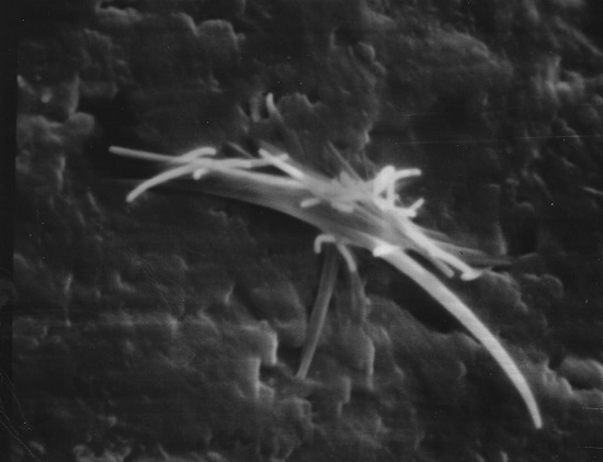
Note: The photo for the header image of this article is from a sample I prepared for my electron microscopy class in 1980.